Academic Staf JPM (Prof Alina)

Associate Professor Dr Tengku Alina binti Tengku Ismail

  • Qualification 
    MD (USM), MCom.Med (Family Health) (USM), PhD (USM)

    Expertise       
    Maternal and women's health, Men's health, Breastfeeding    

    Recognition & Award       

    • Massive Open Online Course (MOOC) on Breastfeeding Self-Learning (Silver Medal) UPM International University Carnival on E-Learning (IUCEL) 2022
    • Anugerah Inovasi Penyusuan Susu Ibu sempena Sambutan Minggu Penyusuan Susu Ibu Peringkat Kebangsaan oleh KKM, 2021
    • Diabelicious Cookies (Gold Medal) 32nd International Invention, Innovation and Technology (ITEX) 2021   
    • MOOC penyusuan susu ibu: pembelajaran kendiri (Gold award), UITM, The Virtual International e-Content Development Competition 2021
    • My Nypa Sugar (Silver Award) (International Conference and Expositions on Inventions by Institutions of Higher Learning, 2019) (Pecipta 2019) 
    • Anugerah Sanggar Sanjung 2019, 2020 & 2021

Supervision (As Main Supervisor)


  • Doctor of Public Health (Active Supervision)

    1. Dr Nionella Stephne Sampil
    2. Dr Nur Hafizah Sukeri
  • Doctor of Public Health (Graduated)

    1. Dr Najihah Mahfuzah Zakria (2019)
    2. Dr Hafizah Ibrahim (2019)
    3. Dr Pathman Arumugam (2020)
    4. Dr Fatin Aina binti Abu Bakar (2020)
    5. Dr Wan Soliha Wan Mohd Hanafi (2022)
    6. Dr Muhammad Zikri Ab Aziz (2023)
    7. Dr Nur Nabila Jusoh (2024)
    8. Dr Tengku Muhammad Fakharuddin Tengku Md Fauzi (2024)
    9. Dr Saidah Adilah binti Mohamed Yusof (2024)

  • Master of Public Health (Active Supervision)

    1. Dr Nur Fazlina Dzulkifli
  • Master of Public Health (Graduated)

    1. Dr Zul Azrizal Salleh (2019)
    2. Dr Siti Fatimah Samsury (2020)
    3. Dr Saidah Adilah Mohamed Yusof (2021)
    4. Dr Nur Dalilah Mohd Zin (2021)
    5. Dr Amalina Asmail@Ismail (2023)
    6. Dr Mohd Fariz Zulrushdi (2024)

Read more …Academic Staf JPM (Prof Alina)

Academic Staf JPM (Prof Ismail)

Associate Professor Dr Mohd Ismail Ibrahim

  • Qualification 
    MD (USM), MComm.Med (Health System Management)(USM) 

    Expertise       
    Health Management focusing on patient quality care and patient safety


    Recognition & Award       
    Anugerah Sanggar Sanjung 2018, 2019, 2020, 2021 & 2022

    Timbalan Pengarah Pusat Rancangan Kokurikulum Kampus Kesihatan
         

Supervision (As Main Supervisor)


  • Doctor of Public Health (Active Supervision)

    1. Dr Mohd Hanif bin Mohd Nawai
    2. Dr Muhammad Solehuddin bin Ishak
    3. Dr Nur Shuhaiza binti Supian
  • Doctor of Public Health (Graduated)

    1. Dr Ahmad Badrurridzwanullah bin Zun (2021)
    2. Dr Afiq Izuddin bin A Rahim (2022)
    3. Dr Ahmad Zulfahmi bin Mohd Kamaruzaman (2022)
    4. Dr Mohamad Hazni bin Abdulah Rahim (2023)
    5. Dr Noorhidayu Monyati binti Mohamed Nor (2023)
    6. Dr Muhammad Iqbal bin Hj Mokhti (2024)

  • Master of Public Health (Active Supervision)

    1. Dr Muamar Iskandar Mohamed Yusoff
  • Master of Public Health (Graduated)

    1. Dr Ahmad Zulfahmi bin Mohd Kamaruzaman (2019)
    2. Dr Ahmad Hazni bin Abd Rahim (2020)
    3. Dr Muhammad Iqbal bin Hj Mokhti (2021)
    4. Dr Muhammad Solehuddin Ishak (2023)
    5. Dr Mohd Hanif bin Mohd Nawawi (2023)
    6. Dr Nur Shuhaiza binti Supian (2024)

Read more …Academic Staf JPM (Prof Ismail)

Academic Staf JPM (Prof Zahir)

Associate Professor Dr Wan Mohd Zahiruddin bin Wan Mohammad

  • Qualification 
    MD (USM), MComm.Med (Epidemiology & Biostatistics) (USM)

    Expertise       
    Epidemiology of Communicable Disease
    Public Health Research on HIV and Tuberculosis
    Research Methodology and Biostastics

    Recognition & Award       
    Ahli Setia Mahkota Kelantan Yang Amat Terbilang (A.S.K) 2023

    Anugerah Perkhidmatan Cemerlang 2010 & 2017
    Head of Community Medicine Department since March 2024  
    Head of Community Family and Case Study Unit 2009-2024
    Alumni of WHO Fellowships Programme on HIV/AIDS Surveillance
    Members of National Coordinating Commitee on HIV

Supervision (As Main Supervisor)


  • Doctor of Public Health (Active Supervision)

    1. Dr Amalina binti Asmail @ Ismail
  • Doctor of Public Health (Graduated)

    1. Dr Sahrol Azmi bin Termizi (2014)
    2. Dr Siti Rohana binti Ahmad (2019)
    3. Dr Abdulloh bin Mazalan (2019)
    4. Dr Mohd Ridzuan bin Mohd Lutpi (2019)
    5. Dr Mohd Shamril bin Mohd Nawi (2020)
    6. Dr Tengku Noor Farhana binti Tengku Khalid, 2022)
    7. Dr Mohd Hazwan bin Baharuddin (2023)
    8. Dr Muhmmad Ikhwan bin Ismail (2023)
    9. Dr Nor Azlina binti Abdullah (2024)
    10. Dr Nur Dalilah binti Mohd Zain (2024)

  • Master of Public Health (Active Supervision)

    1. Dr Sanggary Marimuthu
  • Master of Public Health (Graduated)

    1. Dr Najihah Mahfuzah binti Zakaria (2015)
    2. Dr Siti Rohana binti Ahmad (2015)
    3. Dr Abdullah Mazalan (2016)
    4. Dr Ahmad Hanis bin Ahmad Shushami (2017)
    5. Dr Muhammad Zikri bin Ab Aziz (2019)
    6. Dr Nur Akmal bin Ismail (2020)
    7. Dr Ahmad Syaify bin Berahim (2021)
    8. Dr Mohd Hafiz bin Che Ismail (2021)
    9. Dr Ahmad Syahid bin Ibrahim (2023)
    10. Dr Muhammad Aminuddin bin Che Hashim (2024)

Read more …Academic Staf JPM (Prof Zahir)

ABOUT US

ABOUT US

Background

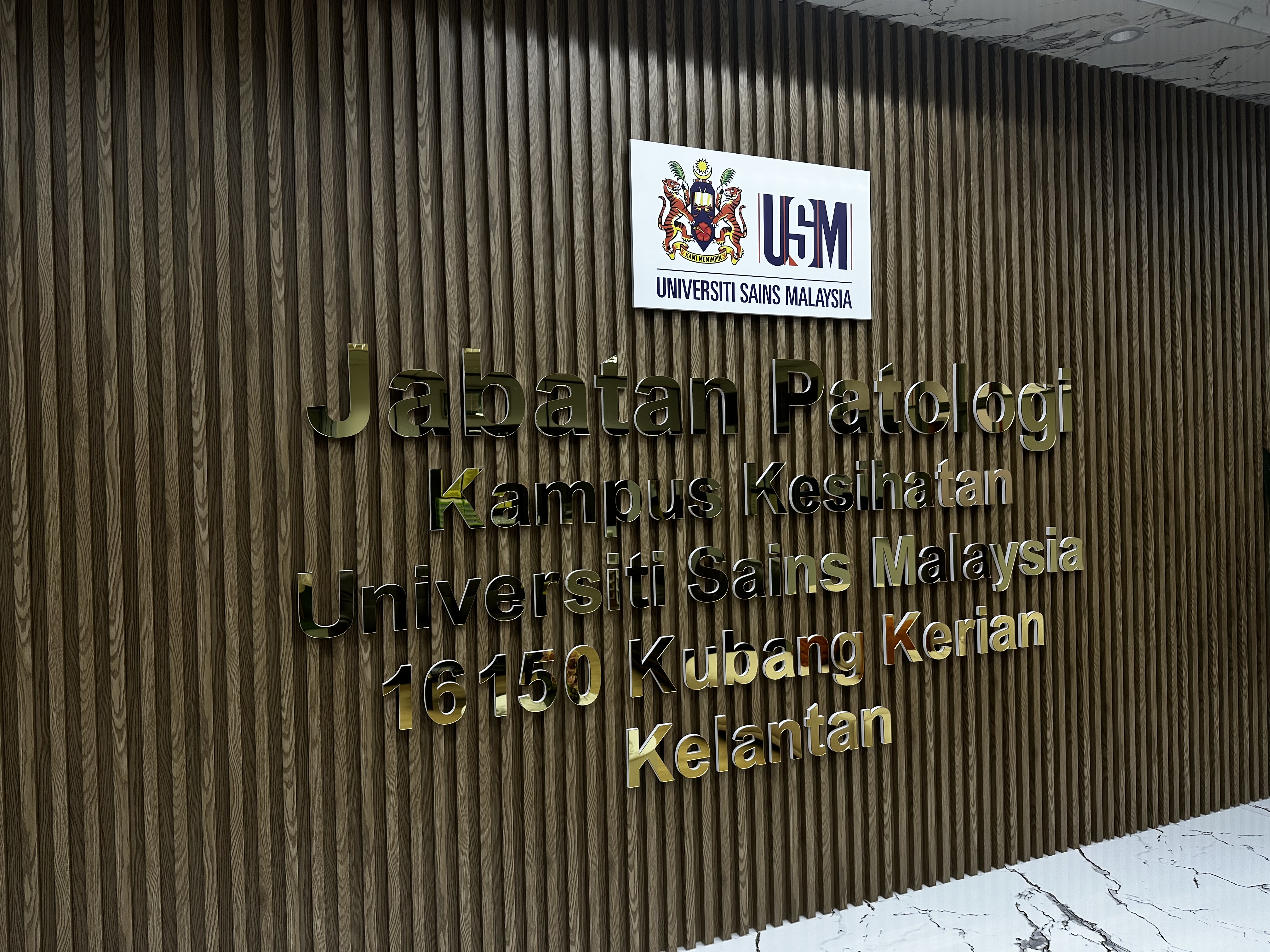
The Department of Pathology was established in 1983, along with the commencement of the Hospital Universiti Sains Malaysia.  It was a big department that provided, basic histo- and cytopathology services, haematology and blood bank services.  Later, the laboratory expanded, providing special staining and immunohistochemical tests. In 1991, a Human Genetic Unit was developed.  However, in 1998, the Haematology Unit and Human Genetic Unit became independent units and were upgraded into a department status, Department of Haematology & Transfusion Medicine, and Human Genome Centre, respectively.

Vision

To be the centre of excellence for the teaching, training and research in pathology.

Mission

The Department of Pathology is dedicated to becoming a premier centre for undergraduate, postgraduate, and subspecialty education. We aspire to lead in clinical pathology and research in Malaysia, distinguished by our commitment to innovation and the integration of evidence-based practices derived from cutting-edge research. Our unwavering goal is to deliver exceptional patient care through excellence in service and scientific advancement.

Head of Department Welcome Note

I would like to welcome you to the Department of Pathology, School of Medical Sciences, Health Campus, Universiti Sains Malaysia. In parallel with our mission and vision, our department has move forward. In the past, many people have no idea what pathologists actually do. They thought the pathology was only part of the undergraduate or pre-clinical subject or just a basic science subjects that need to be memorized, or they immediately think of a doctor performing autopsies. However, they may be connected with pathologists directly or indirectly in many ways: the precise diagnosis of a great deal of diseases are made based on the work of pathologists, and in these cases their function is essential to the successful work of doctors in hospitals.

Today, the pathology has become a very important subject, not only for under-graduates and master of pathology, but also in general hospital services especially surgical based related. Pathological findings often mean the first and foremost step towards recovery. Besides making diagnoses, pathologists support the hospital team (oncologists, surgeons/physicians) by their expertise. We’re the doctors behind the screen.  That is why the department of pathology has undergone a great transformation and expansion throughout its years of service till today.

Other than teaching/ learning activities, we also participate in various research activities and cancer research projects.  In addition, our laboratory has achieved the accreditation and has been recognized as one of the training centers with a good medical laboratory services in accordance to the standards MS ISO 15189:2022. Our aim is to provide an excellent patient care by comprehensive, good quality laboratory testing and patient-focused expert consultation. Patients’ safety is our concern. This mission is supported by continuous quality improvement and leadership in education. Our staff are our most valuable resource and these were achievable with the strength of dedicated staffs working together as a team and one big family.

Our diagnostic services include histopathology with special stain/immunohistochemistry units and cytopathology unit. Frozen section service is also part of our diagnostic services. As a training center for undergraduates and post-graduates, the interesting specimens are prepared in the jar for the learning purposes and kept in the museum. We also provide a link to the pathology museum web site containing images to help the students to understand pathology. The laboratory bench has become our post-graduate training to be future pathologists.  In addition, we have a research laboratory /unit which is dedicated to our research and molecular works. Most of the master of sciences and PhD candidates who involve in pathology work will go through this special unit.  We also have a forensic unit, which was formerly known as mortuary.  Today, the HPE and cytopathology reports are computerized using the Laboratory Information System (LIS), and can be accessed from the wards and clinics. Interestingly, our LIS is customized created in accordance to our MS ISO15189:2022 requirement. Currently, we are working into paperless histo-and-cytopathology reports. 

We aim to expand our services especially in diagnostic molecular pathology, and perhaps one day becomes a cancer center for the east coast of Peninsular Malaysia.  We are also hoping to continue collaborating with our colleagues nationwide to expand our research, teaching and clinical services.

The department has achieved so much in the past and is hoping to achieve more in the future. We are indebted to our fellow clinicians who had contributed so much to the department. We are hoping that this working relationship keeps thriving in the future to come.

May Allah almighty bless the department and guide our path.

Best wishes,

Associate Professor Dr. Faezahtul Arbaeyah Hussain
Head of Department,
Department of Pathology,
School of Medical Sciences,
Health Campus, USM, Kelantan


Read more …ABOUT US

Details of test immuno 5524 2

DETAILS OF TEST


There are 20 diagnostic laboratory tests at our Department described as follows (include tests with MS ISO15189 certified): 

  • 1. Anti-Nuclear Antibody (ANA)*

    Type of specimen: Minimum 3 ml of blood in plain tube.

    Method/Principle: Immunofluorescence (IF) using HEp-20-10 substrate.

    Interpretation: Positive IF (nucleus) at titre 1/80 or more. IF patterns suggest ANA specificity.

    Clinical significance: ANA is a screening test for suspected connective tissue disease. Positive result, especially high titres indicate a possible connective tissue disease. Negative result in 95% to 99% of cases rule out systemic lupus erythematosus (SLE). ANA is also detected (low titre) in other autoimmune diseases such as chronic active hepatitis (CAH), Sjögren’s syndrome, primary biliary cirrhosis (PBC) and rheumatoid arthritis (RA).

    Result: Within 8 working days.


    Note: If ANA is positive, confirm with test for anti-dsDNA, anti-Sm and anti-RNP for SLE [see Anti- Extractable Nuclear Antigens (ENA)].

  • 2. Anti-Double Stranded DNA (dsDNA)*

    Type of specimen: Minimum 3 ml of blood in plain tube.

    Method/Principle: Flourescene Enzyme ImmunoAssay (FEIA) 

    Clinical significance: High titre is suggestive of active SLE, however it is negative in 25% of SLE patients.

    Result: Within 8 working days.

  • 3. Anti-Smooth Muscle Antibody (ASMA)

    Type of specimen: Minimum 3 ml of blood in plain tube.

    Method/Principle: Immunofluorescence (IF) using tissue section as substrate.

    Interpretation: Positive IF at titre of 1/20 or more.

    Clinical significance: High titre of ASMA is detected in patients with autoimmune chronic active hepatitis (CAH) (60-70%), lupoid hepatitis (80%) and other liver diseases including Epstein Barr viral infection.

    Result: Within 10 working days.

    Note: Tests are done together with AMA, LKM and APCA

  • 4. Anti-Mitochondrial Antibody (AMA)

    Type of specimen: Minimum 3 ml of blood in plain tube.

    Method/Principle: Immunofluorescence (IF) using tissue section as substrate.

    Interpretation: Positive IF at titre of 1/20 or more.

    Clinical significance: High titre of AMA is detected in 90% patients with primary biliary cirrhosis (PBC) but also found in other liver diseases (low titre), syphilis, connective tissue disorders and myocarditis.

    Result: Within 10 working days.

    Note: Tests are done together with ASMA, LKM and APCA.

  • 5. Extractable Nuclear Antigens (ENA)*

    Extractable Nuclear Antigen Antibodies (dsDNA, Nucleosomes, Histones, SmD1, PCNA, RPP/PO, SS-A/Ro 60kD, SS-A/Ro 52 kD, SS-B/La, CENP-B, Scl-70, U1-snRNP, AMA M2, Jo-1, PM-Scl, Mi-2 & Ku)

    Type of specimen: Minimum 3 ml of blood in plain tube

    Method/Principle: The test is based on the principle of an enzyme immunoassay. The test strip is composed of a membrane fixed on a plastic support. The intensity of the coloration is directly proportional to the amount of antibody present in the sample.
    Interpretation: A sample is positive for a specific antibody if the colour intensity of the corresponding Antigen Dot is higher than the intensity of the Negative Control Dot. A sample is negative for a specific antibody if the colour intensity of the corresponding Antigen Dot is lower or equal than the intensity of the Negative Control Dot.

    Clinical significance: Anti-Sm is specific for SLE (30-40%), but negative result does not rule out SLE. Anti-RNP is detected in mixed connective tissue diaseses (MCTD) (95-100%) but also in SLE and scleroderma. Anti-SS-A occurs in SLE (25%), Sjogren syndrome (40-50%), congenital complete heart block, cutaneous lupus and is a serologic marker for neonatal lupus. Patients with anti-SS-B most commonly have Sjogren syndrome (23%) and SLE (5-10%) and Sjogren syndrome sicca complex (60%) and neonatal lupus. Anti-Scl-70 occurs in 70% of patients with progressive systemic sclerosis (PSS) and is a specific marker for PSS. Anti-Jo-1 is detected in patients with polymyositis/dermatosytis. Anti-PM-Scl is found in patients with polymyositis and scleroderma. Anti- CENP-A/B is detected in patients with CREST (60%). Anti-PCNA is another specific marker for SLE but is only detected in 5-10% of the patients. Anti-Ku and anti-Mi-2 antibodies are detected in patients with myositis.

    Result: Within 5 working days.

  • 6. Anti-Neutrophil Cytoplasmic Antibody (ANCA)

    Type of specimen: Minimum 3 ml of blood in plain tube.

    Method/Principle: Immunofluorescence (IF) using a monolayer of cells as substrate.

    Interpretation: Positive IF at titer of 1/20 or more.

    Clinical significance: cytoplasmic (c-ANCA) is detected in patients with Wegener’s granulomatosis, microscopic polyarteritis. Perinuclear (p-ANCA) may occur in polyarteritis nodosa, SLE, severe vasculitis and rheumatoid vasculitis.

    Result: Within 10 working days.

  • 7. Anti-Cardiolipin Antibody (ACA)

    Type of specimen: Minimum 3 ml of blood in plain tube.

    Method/Principle: ELISA

    Interpretation: the absorbance values of the samples are multiplied by the Conversion Factor to obtain the anti-cardiolipin antibody concentration in GPL or MPL units.

    Clinical significance: Positive anti-cardiolipin Ab is found in patients with repeated abortions associated with SLE, various venous and arterial thrombotic disorders including cerebral infarction, deep venous thrombosis, thrombocytopenia and pulmonary embolism.

    Result: Within 10 working days.

  • 8. Rheumatoid Factor (RF)

    Type of specimen: Minimum 3 ml of blood in plain tube.

    Method/Principle: Latex agglutination

    Interpretation: Agglutination of latex particles compared to positive control.

    Clinical significance: RF is detected in >80% patients with rheumatoid arthritis (RA). It is also detected in other diseases such as SLE, Sjogren’s syndrome, chronic infections (infective endocarditis & tuberculosis).

    Result: Within 8 working hours.

  • 9. Urine Pregnancy Test (UPT)

    Type of specimen: 20 ml of urine in urine container (preferable early morning urine).

    Method/Principle: Immunoblot (strip).

    Interpretation: Band formation is compared with the control. 2 bands formation indicate positive result.

    Clinical significance: Positive result indicates pregnancy (in pregnancy hCG is increased exponentially for the first 8 weeks, peaks at 10 weeks and start to decline after that until 1/5 of peak level). But very high hCG up to 200,000IU/L suggestive of chrio-epithelioma. hCG can also be detected in other conditions such as persistent trophoblastic diseases, hydatidiform mole, chorioepithelioma of uterus or testis and testicular tumours (monitoring some ovarian and testicular malignancies). False positive UPT can be seen in UTI (urine full of bacteria), proteinuria, hematuria and patient on methadone treatment. False negative UPT maybe found in ectopic pregnancy, toxaemia, foetal death or threatened abortion.

    Result: Within 8 working hours.

  • 10. Anti-Cyclic Citrullinated Peptide Antibody (ACCPA)

    Type of specimen: Minimum 5 ml of blood in plain container.

    Method/Principle: FEIA (Fluorescent enzyme immunoassay).

    Interpretation: Test is considered positive if the antibody titre was greater than 12 IU/ml.

    Clinical significance: Highly specific marker with comparable sensitivity for rheumatoid arthritis compared to RF.

    Results: 10 working days.

  • 11. Allergen-specific IgE Assay*

    Type of specimen: minimum 3 ml blood in plain bottle.

    Method/principle: Patients samples, calibrators and controls are incubated with the allergen. After washing, all these solid phase are incubated with a conjugate. After another series of washing, a flurogenic substrate is incubated with these solid phase and then stop solution is added. The end points are read by fluorometry. The standard curve is generated. Calibration data are fit and control values are interpolated on the standard curve.

    Clinical Significance: Assessing the level of allergen specific IgE in a Patients’ serum in conjunction with a clinical evaluation based on patient history. This will help the clinician to confirm a diagnosis of allergy and assist in the treatment of the patient.Result: within 5 working days.

    Result: Within 8 working days

  • 12. Skin Antibodies Panel

    Type of specimen: Minimum 3 ml of blood in plain tube.

    Method/Principle: Immunofluorescence (IF) using Dermatology Mosaics 7.

    Interpretation: Fluorescence pattern (positive reaction). Antibodies against spine cell desmosomes react with the surface antigens of keratinocytes.


    Tissue sections of oesophagus and tongue show a characteristic, honeycomb-like, smooth fine granular fluorescence of the intercellular substance, which is mainly spread over the entire stratum spinosum.


    Autoantibodies against epidermal basement membrane show a fine-linear staining between the stratum basale and the connective tissue.


    Antibodies against plakins react with the transitional epithelium (basement membrane and desmosomes) of rat urinary bladder. Antibodies against epidermal basement membrane cause a fine linear staining between the stratum basale and the connective tissue. Additionally, a reaction of the intercellular substance of the transitional epithelium can be visible (antibodies against desmosomes).


    Antibodies against basement membrane structures react with salt-split skin. Anti-BP180, anti-BP230 and anti-LAD97 cause a staining of the epidermal side, while antibodies against laminin 332, collagen VII and other antigens stain the dermal side of salt-split skin.


    Antibodies against laminin 332 (LAM332, formerly laminin 5) react specifically with the transfected cells of the test substrate. They cause a smooth to finely granular cytoplasmic fluorescence, in part with slightly pronounced cell membranes. The cell nuclei are usually not or only slightly stained.


    Antibodies against BP230gC and collagen VII show a smooth to finely granular cytoplasmic fluorescence in the transfected cells of the respective test substrate, partly with fluorescence of the cell membrane. The cell nuclei are usually not or only slightly stained.


    Antibodies against desmoglein 1 and desmoglein 3 show a smooth to finely granular cytoplasmic fluorescence in the cells of respective test substrate, partly with slightly pronounced cell membranes. The area of the cell nucleus is usually not or only slightly stained.


    Clinical significance: Autoimmune bullous dermatoses belong to the organ-specific autoimmune diseases. They are characterised by the formation of autoantibodies against structural proteins of the skin. These structural proteins establish the cell-to-cell contact in keratinocytes within the epidermis and the adhesion of the epidermis to dermis. Autoimmune bullous dermatoses are divided into 4 groups based on their target antigens and the localisation of the blisters: pemphigoid diseases, including epidermolysis bullosa acquisita, dermatitis herpetiformis, pemphigus diseases and paraneoplastic pemphigus.  

    Result: Within 4 months.

  • 13. HLA Typing Test (Transplantation)


    Type of specimen: minimum 3 ml x 3 tubes in EDTA bottle

    Method/Principle: Extraction of genomic DNA from the mononuclear cells followed by  low-resolution  sequence-specific primer amplification (PCR- SSP).Primer pairs are designed to have perfect matches only with a single allele or group of alleles.  The perfectly  matched primer pairs result in the amplification of target sequences while mismatched primer pairs do not result in amplification.

    Interpretation: The presence or absence of appropriately sized bands will be assessed and analyzed using worksheet.  The results of molecular typing were converted to the

    serologically   stablished  HLA  phenotypic equivalents.

    Clinical Significance: Pre-requisite for organ especially bone marraw transplantation in order to find matched donor.   Tissue typing is done for HLA class I (HLA-A, HLA-B, HLA-C) and class II  (HLA-DR, HLA-DQ).

    Result:  3 months

    Note: An appointment with the Laboratory is essential for this test. Blood sample of donors and recipient need to be send together.   For donors HLA class II wil be typed first, if DR locus is matched, then  proceed for HLA class I

  • 14. TBNK for HIV Positive & Immunodeficiency*

    Type of specimen: paediatric - 1 ml of fresh anti-coagulated blood in EDTA container; adult – 3 ml fresh anti-coagulated blood in EDTA container.

    Method/Principle: Flow cytometry using monoclonal antibody against various lymphocyte subpopulations.

    Interpretation: Percentage of cells with positive staining.

    Clinical significance: Lymphocyte immunophenotyping is mainly done to support diagnosis of AIDS/HIV infection (low CD4 population (<200 cells/ul of blood is characteristic of AIDS). Lymphocyte counts are also useful for B & T cells deficiencies.

    Result: Within 8 working hours. 

    Note: An appointment with the Laboratory is essential for this test (at least one day earlier). All  specimen must arrive at Immunology Laboratory before 12pm and within 4 hours of collection. 

  • 15. T Cell Proliferation Test (CFSE; for SCID diagnosis)

    Type of specimen: Suspected patient: At least 2 ml of fresh blood in sodium heparin tube (green-top) AND healthy control (at least 2 ml of fresh blood in sodium heparin tube)

    Method/Principle: Flow cytometry (CFSE)

    Interpretation: Percentage of T cells that proliferate in the suspected patient compared with healthy control samples. 

    Clinical significance: Severe combined immunodeficiency (SCID) and other primary immunodeficiency disorders. 

    Results: Within 7 working days.

    NoteAn appointment with the Laboratory is essential for this test (at least one day earlier). All specimen must arrive at Immunology Laboratory before 10am and within 24 hours of collection.


  • 16. HLA Typing Test (Disease Association- B27)

    Type of specimen: minimum 3 ml x 3 tube of blood in EDTA bottle.

    Method/Principle: Extraction of genomic DNA from the mononuclear cells followed by  low-resolution  sequence-specific primer amplification (PCR- SSP).Primer pairs are designed to have perfect matches only with a single allele or group of alleles.  The perfectly  matched primer pairs result in the amplification of target sequences while mismatched primer pairs do not result in amplification.

    Interpretation: The presence or absence of appropriately sized bands will be assessed and analyzed using worksheet.  The results of molecular typing were converted to the

    serologically   stablished  HLA  phenotypic equivalents.

    Clinical Significance: The human leukocyte antigen HLA-B27 is strongly associated with development of a group of inflammatory arthritides collectively known as the spondyloarthritides, a group of related diseases including ankylosing spondylitis and reactive arthritis.  It can precede the onset of ankylosing spondylitis by approximately three years

     Acute anterior uveitis, are also highly correlated with possession of HLA-B27. With lower frequency, the presence of the HLA-B27 allele has correlated with inflammatory bowel disease, psoriatic arthritis, and reactive arthritis.

    Result:  3 months

    Note: An appointment with the Laboratory is essential for this test. Blood sample of donors and recipient need to be send together.  

*NOTE :  MS ISO 15189 CERTIFIED.


1. Inquiries regarding Immunological Lab Tests can be forwarded to our Immunopathologist:
  • Dr. Nurul Khaiza Yahya (This email address is being protected from spambots. You need JavaScript enabled to view it.; ext. 6224)
  • Dr. Noor Suryani Mohd Ashari (This email address is being protected from spambots. You need JavaScript enabled to view it., ext. 6225)
  • Dr. Wan Majdiah Wan Mohamad (This email address is being protected from spambots. You need JavaScript enabled to view it. ext. 5851) 
  • Dr. Nur Diyana Mohd Shukri (This email address is being protected from spambots. You need JavaScript enabled to view it., ext. 6222)
  • Dr. Syazwani Mohd Tajudin (This email address is being protected from spambots. You need JavaScript enabled to view it., ext. 6227)
2. Inquiries regarding T Cell Proliferation Test (CFSE) can be forwarded to our Laboratory Immunologist:
  • Assoc. Prof. Dr. Wong Kah Keng (This email address is being protected from spambots. You need JavaScript enabled to view it.; ext. 6226)
  • For appointment setting, please contact the Medical Laboratory Technologist in duty (Flow cytometry team) at Immunology Lab counter 09-767 4000

QUICK LINK

6.09947385,102.28285600705948

Read more …Details of test immuno 5524 2